Author: Ken Stauver

  • XRD tube bisection

    XRD tube bisection

    XRD tubes rely on the ability to precisely regulate the flow of electrons from the filament to the anode in order to create x-ray emissions. That requires a completely evacuated envelope to avoid having the high-voltage short to ground. The resulting symptom is that the high voltage generator will shut down almost instantly when the high-voltage potential is applied.

    This particular tube looked fine on the outside and didn’t even look all that old so it was a little surprising to find it behaving like the vacuum in the envelope had been compromised. However, after two tests, it was definitively bad so it was set aside for disposal. The first step is simply to remove the head of the tube which is little more than a mounting flange and cooling water distribution device, but when it was removed, the back side of the anode is cooled directly with water and showed some of the most extreme pitting we’d ever seen!

    The next step is to break the ceramic envelope off the metal body of the tube. It was full of water which was obviously the cause of the problem. The pitting must have broken through to the vacuum envelope, which then sucked in water until it was 100% full. Hence the lack of any “sloshing” while we handled the tube.

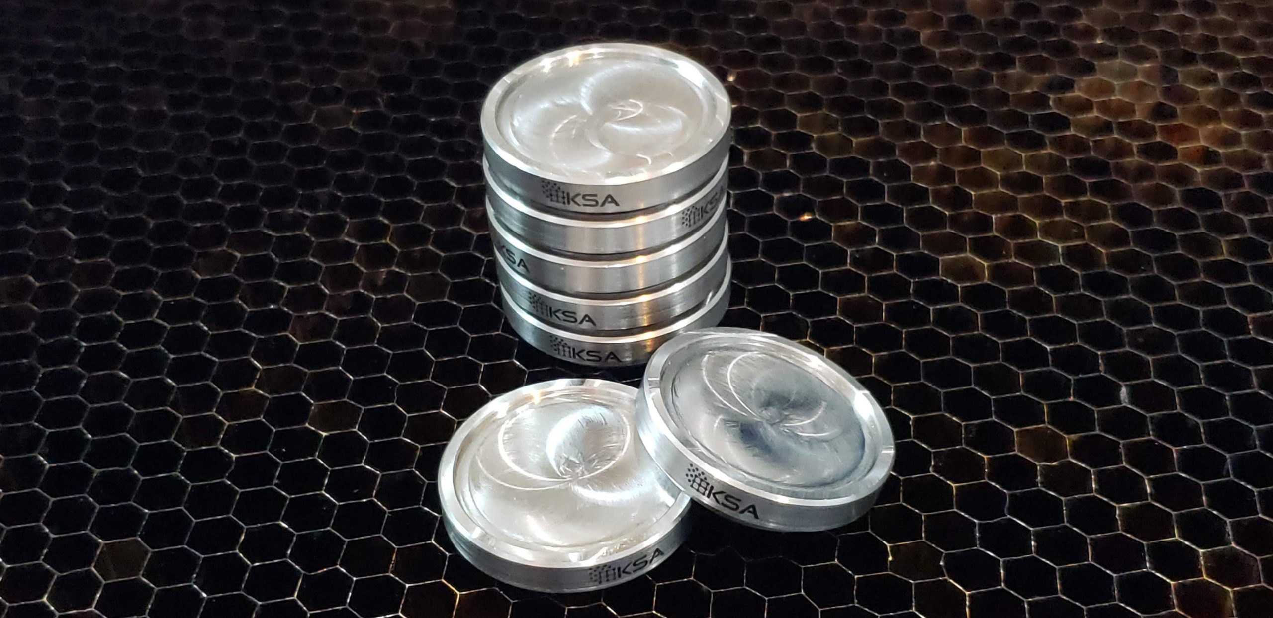
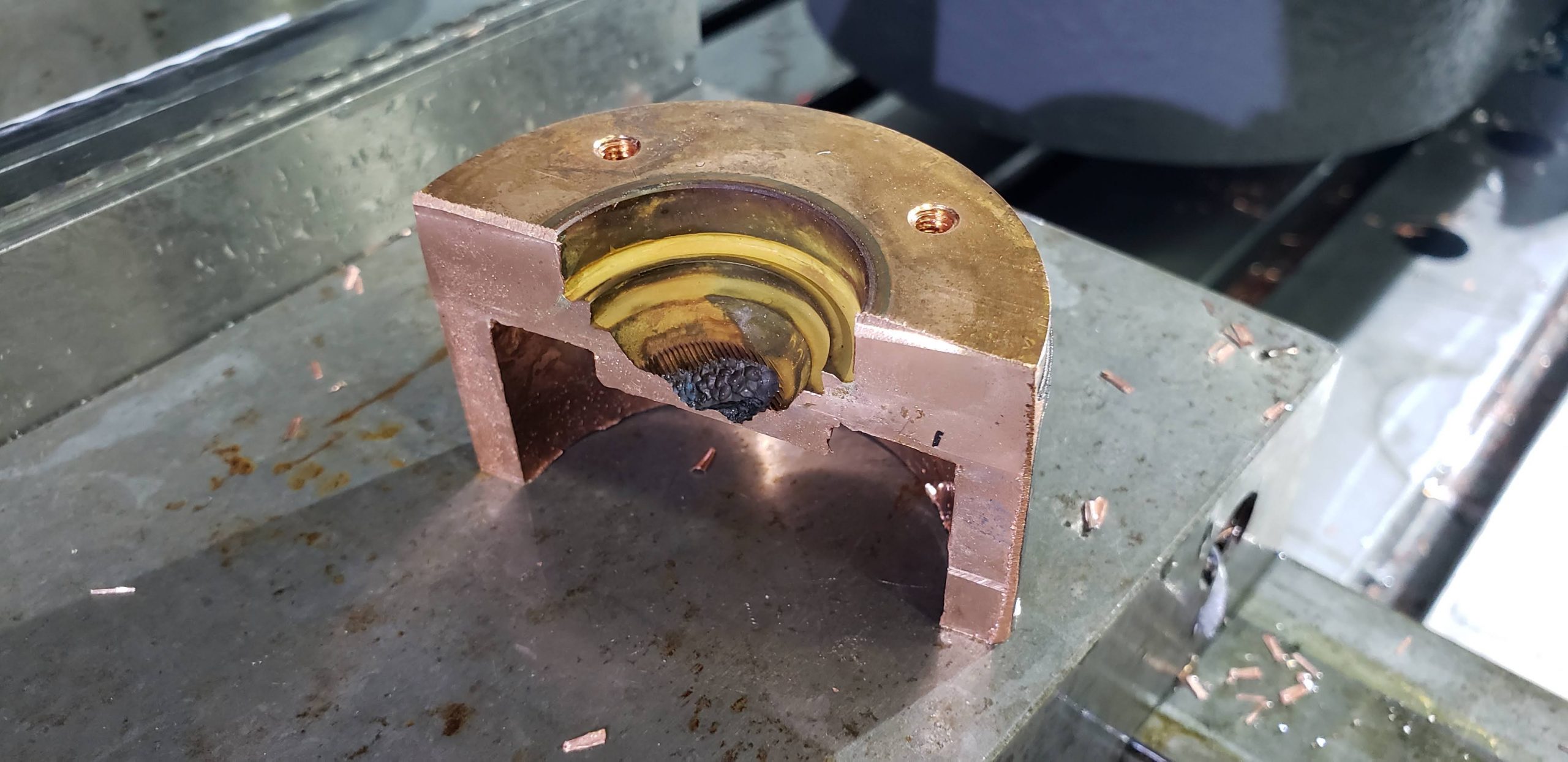
    Cross sectioning the tube anode showed just ow extensive the pitting was. The actual hole was so small that I could only guess where it had been, but it was definitely there.

    We disassemble the x-ray tube on XRD systems during every preventative maintenance procedure and definitely would have caught this before it failed completely. It was most likely caused by either very low quality tap water being used in the recirculating chiller or some additive designed to prevent scale or algae.

  • Mystery crystals

    Mystery crystals

    Curiosity may have killed the cat, but it’s the lifeblood of a well-functioning analytical lab. A few days ago, Todd was preparing a water chiller for shipping and washed out some corrosion products with acetic acid. The resulting solution was left in a beaker over the weekend and when we returned, he noticed that it had formed rather large crystals. So he did what any curious person with a lab full of XRD instrumentation would do. He ground it up into a fine powder and ran it through a D4 Endeavor. The resulting pattern was definitive and made complete sense given the brass metals involved in the corrosion product and the acetic acid solution. 

    The data was imported into MDI Jade Pro for phase identification. Jade makes it very easy to take the analysis all the way through whole pattern fitting (WPF/Rietveld) when paired with the COD or ICDD PDF-4 databases. I generated a full report even though the data was not nearly the quality we would usually require for this type of analysis for a client.

  • PMMA vs Aluminum sample holders

    PMMA vs Aluminum sample holders

    Plastic sample holders have been the default option XRD for decades. They’re inexpensive to make, good enough for most purposes, and very resistant to a wide range of chemicals. Seems like a “win” all around right? As long as they’re made correctly and from the proper materials, these work just fine. Spoiler alert: 3D printed thermoplastics have a distinct structure so if you try making your own, be sure they’re well out of the irradiated area.

    We’ve seen a big move toward more zero-background holders in the last few years and they’re definitely incredible, but many XRD users aren’t concerned with running extremely small volumes as much as they are eliminating or minimizing the scattering of x-ray by the plastic at low angles. As a general rule for polycrystalline materials, the lower the average atomic number of the material, the more efficiently it scatters x-rays. We’re not talking about Bragg diffraction. More like shining a flashlight on a white sheet of paper. Compton and Raleigh scattering are the two effects at work here.

    We use this effect to great advantage in the XRF world when we want to check out the emission spectrum of the x-ray tube. Running a piece of graphite as the sample allows us to collect a scan which shows all the different energies in the emission spectra. Unfortunately, scatter is highly problematic in XRD.

    This is why we supply aluminum holders with our refurbished systems and why we use mostly aluminum holders at Texray. Different plastics can perform differently, but the problem is characterized by a wide, low hump centered around 10 degrees 2Theta and with a width of about 10 degrees. This is a key area in many materials so anything we can do to clean up the background is usually beneficial.

  • What’s really coming out of my XRD tube?

    What’s really coming out of my XRD tube?

    We’ve been experimenting with better ways to quantify the quality of XRD tubes in the shop. We use these tests on new and used tubes to monitor performance in two key areas. 1) Intensity 2) Spectral purity.

    What we’ve settled on is a test that involves a wavelength-dispersive approach which gives us a lot of intensity to work with while eliminating background scatter and fluorescence effects. Basically, we’re able to extract more information from the data because the “noise” is almost zero.

    We used Jade Pro to evaluate the scans, but they’re not D-spacing vs intensity as one would normally expect. This scan represents Wavelength vs intensity more like one would see in a WDXRF spectrometer. Cu KA1 and 2 are obvious, as is Cu KB1. Many of the current generation of XRD users have never seen a W LA1 peak in their data, but it’s clearly visible here as this is an older tube. What I’ve never been able to see before is the W La2 peak in the green scan. You’re looking at a peak that is ~62eV separated from W La1. No XRD detector on the market has energy resolution like that so these would always be lumped together so you’d see a series of additional peaks from every d-spacing in the sample in the diffractogram. Only a handful of detectors (our SDD-150 for example) could even separate the W La from the Cu K lines. That’s the power of wavelength-dispersive techniques. Incidentally, the most common device for cleaning up superfluous energy emissions in XRD data is a diffracted-beam monochromator and they eliminate all the W La through a secondary diffraction event much like what we’re doing.

    Characterizing emissions is nothing new. In fact, I started wanting to improve this after listening to a talk at DXC about Jim Cline’s famous XRD system which is used at NIST to perform the primary data collection on the CRMs we all use. To paraphrase Sir Arthur Conan Doyle, “When you explain every extraneous data point, the remaining information is the pure truth of the sample”.

  • Bruker D2 Phaser Sample Holders

    Bruker D2 Phaser Sample Holders

    KS Analytical Systems now offers custom sample holders for the Bruker D2 Phaser 6-position autosampler. These can be finished to order with any depth and diameter of well or a zero-background plate (ZBH) with or without a well ground in the surface.

  • Fully digital autosampler install

    Fully digital autosampler install

    Another fully-rebuilt, digital autosampler out in the wild. This one is on a system that already has one of our Si-Drift Detectors and an awesome ICDD Jade Pro/PDF-4+ software package. We’ve got all the fancy new hardware at our in-house lab, but when we need the absolute best data, this is our goto configuration.

  • Sample holders for Rigaku Miniflex systems

    Sample holders for Rigaku Miniflex systems

    KS Analytical Systems now offers custom sample holders for Rigaku Miniflex systems. The 6-position autosamplers and rotation stages (and even some fixed stages) make use of the magnetic disk design for holding powder without taking up much extra room in the diminutive benchtop.

    • Top-loading
    • Rear-loading
    • Zero-background
  • Energy-dispersive detector systems for XRD applications

    Energy-dispersive detector systems for XRD applications

    Energy resolution

    • 140eV under ideal conditions.
    • All KB peaks eliminated electronically.
    • W LA1 (8.40 KeV) lines eliminated from Cu KA1,2 (8.04 KeV) scans even with thoroughly contaminated tubes.
    • Common fluorescence energies (i.e. Fe when Cu tube anodes are used) are greatly reduced. (Brehmstralung effects are impossible to remove completely)
    • Most PSD detectors offer no better than 650eV. This allows for a great deal of fluorescence energy to pass as well as W LA1 from older Cu tubes.

    Low angle scatter

    • The detector mounts in place of the traditional scintillation counter allowing for use of automated variable (motorized) or interchangeable aperture slits to control angular resolution. Scans starting from 0.5 degrees 2? are possible with the proper slit arrangement just as they are with the scintillation counter. The user controls the intensity vs angular resolution of the scan based upon the ideal conditions for their work rather than the limitations of the hardware.
    • Position sensitive detectors are wide open by design which necessitates knife edges over the sample and additional mechanical aperture plates to block air scatter at low angles. Closing off the detector limits the useable channels and reduces the benefit of these detectors dramatically.

    Truly zero maintenance design

    • No delays – The detector is ready to collect data almost as soon as power is applied.
    • No external cooling – Air backed Peltier cooling eliminates the need for water circulation and/or liquid nitrogen.
    • Zero maintenance vacuum design eliminates reliance on an ion pump/backup battery.
    • 12-month warranty against hardware failure under normal use.

    Versatility

    • The Digital Pulse Processor (DPP) includes a usb interface allowing for adjustment and refinement should they be necessary for a particular application. With optional software, full quantitative EDXRF analysis can be performed.

    The detector can be set for any common XRD anode (energy) easily. Multiple energies may even be configured to allow for use with various anodes without the need for additional hardware. We specialize in Siemens (now Bruker) XRD and WD-XRF instrumentation and have installation kits ready for the D500, D5000 and D5005. The output is a standard BNC cable with a 5V square pulse output which is standard across every manufacturer we’ve worked with. Kevex and Thermo Si(Li) detectors used this same output.

    Please contact KS Analytical Systems for a quote.

  • Texray Laboratory Services

    Texray Laboratory Services

    Texray Laboratory Services provides XRD and WDXRF based analysis services including corrosion analysis, quantitative and qualitative material composition analysis, mineralogical (geological) analysis, industrial hygiene measurements and many other advanced XRD techniques (non-ambient temperature and thin-film measurements). Learn more by visiting the Texray-Lab homepage at www.texray-lab.com.